History, Examination and Assessment of Pain course


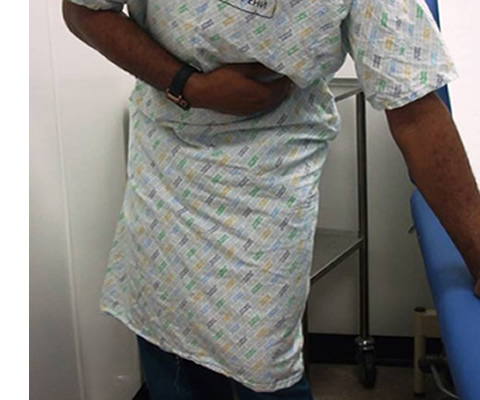
This session describes the factors that must be taken into account when examining and assessing a patient who is in pain.
Learning Objectives
By the end of this session you will be able to:
- Take a patient's pain history
- Identify the psychological factors involved in pain
- Differentiate between nociceptive and neuropathic pain
- Choose the appropriate assessment tool to measure pain
Since all healthcare workers deal with patients who have either transient or more permanent pain, it is important that all team members are aware of not only their own responsibility for assessing and monitoring patients' pain, but also the remit of other team members.
Senthil is a final year registrar in Anaesthesia and Pain medicine in London. His special interests include management of neuropathic pain and promoting education of all healthcare professionals in pain assessment and management.
He is the Secretary of British Pain Society Special Interest Group, Pain in Developing Countries, and organizes workshops and promotes pain management in these parts of the world.
Senthil is a content author for the e-PAIN Project.


- Radiology – Integrated Training Initiative (R-IT...
- Posted By eIntegrity Healthcare e-Learning
- Posted Date: 2025-01-10
- Location:Online
- This session reviews which chambers contribute to the ‘normal cardiac outline’ and examines how specific chamber enlargement or absence alters the normal cardiac shape. It will describe the different diagnoses, which result in characteristic
- Radiology – Integrated Training Initiative (R-IT...
- Posted By eIntegrity Healthcare e-Learning
- Posted Date: 2025-01-10
- Location:Online
- This session provides an overview of the clinical manifestations of haemangiomas, lymphatic and vascular malformations, their radiological work-up plus typical imaging findings.
- Radiology – Integrated Training Initiative (R-IT...
- Posted By eIntegrity Healthcare e-Learning
- Posted Date: 2025-01-10
- Location:Online
- This session covers imaging and diagnosis of paediatric mediastinal masses, and is organised based on their location in the mediastinum.
- Radiology – Integrated Training Initiative (R-IT...
- Posted By eIntegrity Healthcare e-Learning
- Posted Date: 2025-01-10
- Location:Online
- This session discusses radiographic anatomy of the normal mediastinum and its compartments containing different structures and why this is useful when interpreting an abnormal image. The silhouette sign and how it is used to localise a mass on chest radio
- Radiology – Integrated Training Initiative (R-IT...
- Posted By eIntegrity Healthcare e-Learning
- Posted Date: 2025-01-10
- Location:Online
- The session looks at pneumothorax, pneumomediastinum, air leaks in neonates, air leaks in older children and post-traumatic air leaks.


